

What is time-lapse embryo monitoring?
Time-lapse embryo monitoring is a continuous imaging technology built directly into IVF incubators. Integrated cameras capture thousands of images of developing embryos at intervals ranging from 5 to 20 minutes, all without removing the embryos from their controlled environment. Those images are then compiled into video sequences that show the full arc of embryo development from fertilization through blastocyst formation.
The core advantage is observation without disturbance. Traditional IVF requires embryologists to physically remove embryos from the incubator at least once daily to check their development under a microscope. Every removal briefly exposes the embryo to changes in temperature, gas composition, and handling. Time-lapse systems eliminate that disruption entirely.

What makes this technology clinically meaningful is the type of data it generates. Rather than a single daily snapshot, embryologists gain access to what researchers call morphokinetic data: the precise timing of cell divisions, pronuclear appearance and fading, blastomere symmetry, and blastocyst formation. These dynamic details are simply invisible in conventional daily checks.
Key features of time-lapse embryo monitoring at a glance:
- Continuous imaging at 5–20 minute intervals inside the incubator
- No embryo removal required for assessment
- Full video record of embryo development from fertilization onward
- Morphokinetic data capture: cell division timing, symmetry, pronuclear events
- Software-assisted annotation and embryo ranking tools
- Stable culture environment maintained throughout the observation period
How does time-lapse monitoring compare to traditional embryo culture?
The practical differences between these two approaches go beyond just the technology used. They reflect fundamentally different philosophies about how embryos should be observed and assessed during the IVF cycle.
In conventional IVF, embryologists remove embryos from the incubator once a day, place them under a light microscope, assess the number and appearance of cells, and return them to the incubator. The assessment is accurate for that single moment in time, but it misses everything that happened between checks. Cell division events that occur and resolve within hours, irregular cleavage patterns, or early signs of fragmentation can go entirely undetected.
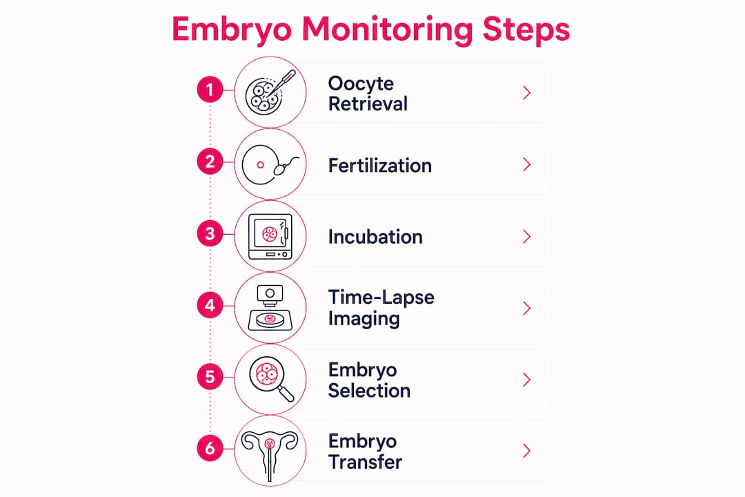
Time-lapse culture keeps embryos inside the incubator continuously. Assessment happens through a screen connected to the integrated camera system, at any point during the culture period, without any physical handling. The embryologist reviews a dynamic video record rather than a static image.

| Feature | Traditional embryo culture | Time-lapse monitoring |
|---|---|---|
| Embryo removal for assessment | Daily | Never |
| Observation frequency | Once per day | Every 5–20 minutes |
| Environmental stability | Interrupted briefly each day | Continuous |
| Data type | Static morphology snapshot | Dynamic morphokinetic record |
| Embryo ranking basis | Appearance at one time point | Developmental timing and behavior |
| Embryologist workflow | Hands-on daily checks | Remote screen-based review |
Several practical distinctions are worth noting for patients:
- Environmental consistency: Closed time-lapse systems maintain temperature, oxygen levels, and CO₂ concentration without interruption, which may support embryo viability.
- Workflow flexibility: Embryo assessment at any time during the day becomes possible, reducing the pressure of a single daily check window.
- Richer data: Transient developmental events that last only minutes, such as irregular cell divisions, are captured on record rather than missed between checks.
- Cost: Time-lapse systems add meaningful cost to an IVF cycle, which varies by clinic and is not uniformly covered by insurance.
How do time-lapse incubators actually work?
The technology inside a time-lapse incubator is more specific than the term suggests. Understanding the hardware helps patients ask better questions when evaluating clinics.
Two distinct system types are used in clinical practice. The first is an open system, where an optical camera unit is placed inside a conventional box incubator. The second is a closed system, a fully integrated bench-top incubator with a built-in inverted microscope and digital camera. Closed systems are more common in modern IVF labs because they combine optimized culture conditions with continuous imaging in a single unit.
Here is how the imaging process works in practice:
- Embryos are placed in specially designed culture dishes specific to each system’s format.
- An inverted microscope within the incubator captures digital images at multiple focal planes, typically every 5–15 minutes throughout the culture period.
- Images are processed automatically by the system’s software into a time-lapse video sequence covering the full development period.
- Embryologists review the video on an external monitor, annotating key developmental events using integrated software tools.
- Annotation data feeds into scoring algorithms that rank embryos based on their morphokinetic profiles.
Pro Tip: Not all time-lapse systems use the same algorithms or scoring criteria. When speaking with your clinic, ask specifically which system they use and whether their algorithm has been validated in their own patient population, since results from one system are not always directly comparable to another.
The software layer is where significant variation exists between clinics. Some use the algorithm provided by the system manufacturer. Others have developed in-house selection models tailored to their specific patient population and laboratory conditions. This variation is one reason why published outcomes from time-lapse studies can differ substantially across institutions.
How embryologists assess embryos using time-lapse imaging
Embryo assessment with time-lapse technology centers on morphokinetics: the study of how an embryo develops over time, not just what it looks like at a single moment. This is where the technology offers its most distinctive clinical value.
Embryologists annotate specific developmental milestones as they appear in the video record. The key parameters include:
- Pronuclear appearance and fading (tPN, fPN): The timing of when the two pronuclei appear after fertilization and when they fade before the first cell division.
- First cleavage (t2): The exact time at which the fertilized egg divides into two cells.
- Subsequent cleavage timings (t3, t4, t5…): The timing of each subsequent cell division through the cleavage stage.
- Cell cycle intervals: The duration between successive divisions, which correlates with developmental competence.
- Blastomere symmetry: Whether dividing cells are equal in size, an indicator of normal development.
- Blastocyst formation timing (tB): When the embryo reaches the blastocyst stage, a key milestone before transfer or freezing.
The clinical significance of this data lies in what static daily assessments cannot capture. Transient events, such as an irregular three-cell stage that resolves before the daily check, or a brief period of fragmentation that later compacts, are permanently recorded in the time-lapse video. Whether those events reliably predict implantation success is a separate question, addressed in the evidence section below.
What does the clinical evidence say about time-lapse embryo selection?
Clinics that use time-lapse systems often apply selection algorithms to rank embryos before transfer. These algorithms assign scores or categories to embryos based on their morphokinetic profiles, with the goal of identifying the embryo most likely to implant successfully.
The evidence on whether those algorithms actually improve outcomes is more nuanced than the technology’s appeal might suggest.
Key findings from the clinical literature:
- Randomized controlled trials show that time-lapse imaging does not improve live birth rates compared to traditional incubation for most IVF patients.
- The UK’s Human Fertilisation and Embryology Authority (HFEA) classifies time-lapse as a treatment add-on, advising patients to discuss expected individual benefits with their clinic before paying for it.
- The predictive power of current morphokinetic algorithms remains limited, with AUC levels below 0.8 in published studies, indicating moderate rather than strong predictive accuracy.
- Existing algorithms are largely built on annotations at the same few traditional time points used in conventional assessment, meaning the vast majority of time-lapse data remains unused.
- Despite mixed outcome data, time-lapse systems do provide a stable culture environment and generate large datasets that may support future algorithm development.
What this means for patients: Time-lapse monitoring offers real observational and environmental benefits. The technology itself is well-validated as a safe and effective way to culture embryos. The question of whether the selection algorithms built on top of it improve your specific chances of pregnancy is where the evidence remains unsettled.
The HFEA’s position reflects a broader consensus in reproductive medicine: the technology is useful and safe, but patients should not assume it will directly increase their probability of a live birth. Clinics that present time-lapse as a proven success-rate booster are overstating what the current evidence supports.
Where is time-lapse embryo technology headed?
The most significant development on the horizon is the application of machine learning to time-lapse image datasets. Current algorithms analyze a small fraction of the data each imaging session generates, focusing on the same developmental milestones embryologists have tracked manually for decades.
Machine learning approaches work differently. Rather than annotating specific time points, these systems analyze every image in the sequence, searching for subtle visual patterns that correlate with implantation and live birth outcomes. The goal is to extract predictive signals that human annotation has never captured.
Several directions are actively being explored:
- Integrated multi-modal analysis: Combining time-lapse morphokinetic data with genetic information from preimplantation genetic testing (PGT) and metabolic markers from spent culture media.
- Standardized algorithm development: Building algorithms that perform consistently across different labs and patient populations, rather than being optimized for a single clinic’s conditions.
- Remote embryo assessment: Time-lapse systems already permit embryo review from outside the laboratory. Expanding this capability could support centralized quality review and specialist consultation.
- Embryologist training tools: The video record from time-lapse systems provides an unmatched resource for training embryologists to recognize developmental patterns, including rare or abnormal events.
The ethical dimension of AI-assisted embryo selection deserves patient attention. As algorithms become more powerful, questions arise about transparency: patients and clinicians should understand what criteria a system uses to rank embryos, and whether those criteria have been validated in populations similar to the patient being treated. Clinics adopting new AI tools carry a responsibility to communicate their validation status clearly.
How to navigate time-lapse monitoring with your fertility clinic
Understanding what time-lapse monitoring is and what the evidence shows puts you in a much stronger position when discussing it with your care team. The decision about whether to use it involves clinical, financial, and personal factors that vary by individual.
Evaluating whether a clinic offers time-lapse technology:
- Ask specifically which time-lapse system the clinic uses and whether it is an open or closed system.
- Ask whether the clinic uses the manufacturer’s algorithm, an in-house algorithm, or no algorithm at all.
- Request information on how many embryo transfers the clinic has performed using time-lapse selection, and whether they track outcomes separately.
Questions about clinical benefit and eligibility:
- For which patients does the clinic recommend time-lapse monitoring, and why?
- Does the clinic have data from their own patient population showing whether time-lapse selection has improved outcomes?
- Are there specific clinical situations, such as repeated implantation failure or poor embryo development history, where time-lapse monitoring may offer more targeted benefit?
Cost and insurance considerations:
- Time-lapse monitoring typically adds cost to an IVF cycle. Ask for the specific fee before committing.
- Most U.S. insurance plans do not cover time-lapse as a standalone add-on. Check your policy’s language around “experimental” or “investigational” procedures.
- Review Life IVF Center’s insurance coverage options to understand what financial support may be available for your treatment plan.
Patient counseling and informed consent:
- Any clinic offering time-lapse monitoring should provide clear written information about what the technology does and does not do.
- Patients have the right to understand the evidence base, including the HFEA’s classification of time-lapse as an add-on with mixed evidence for success rate improvement.
- Counseling during IVF can help you process the emotional weight of these decisions alongside the clinical information.
Pro Tip: Before agreeing to time-lapse monitoring as a paid add-on, ask your embryologist to explain specifically how the technology would change the embryo selection decision in your case. If the answer is that it provides additional data but may not change which embryo is transferred, that context helps you weigh the cost against the benefit honestly.
What you should take away about time-lapse embryo monitoring
Time-lapse embryo monitoring is a well-established, safe technology that offers genuine observational and environmental advantages over conventional daily checks. Its clinical benefit for improving live birth rates, however, remains an open question that patients deserve to understand clearly before making financial decisions.
Core points to carry forward:
- The technology works as described. Continuous imaging inside a stable incubator, without embryo removal, is a real and validated improvement over daily manual checks.
- Morphokinetic data is richer than static assessment. Time-lapse captures developmental events that conventional monitoring misses entirely.
- The evidence on outcomes is mixed. Randomized controlled trials have not consistently shown improved live birth rates, and the HFEA classifies this as a treatment add-on with uncertain clinical benefit for most patients.
- Algorithm quality varies by clinic. The scoring system a clinic uses matters as much as the imaging technology itself, and not all algorithms are equally validated.
- The future is promising but not yet realized. Machine learning and integrated multi-modal analysis may substantially improve embryo selection accuracy, but those advances are still in development.
- Patient empowerment starts with informed questions. Knowing what to ask your clinic, and understanding the honest limits of current evidence, puts you in the best position to make decisions that align with your goals and circumstances.
For patients exploring how IVF technology fits into their broader treatment plan, time-lapse monitoring is one piece of a larger picture. Understanding the signs of healthy embryo development, the full IVF cycle, and how your clinic approaches embryo selection gives you the clearest view of your options.
Explore personalized IVF care at Life IVF Center

Life IVF Center’s Precision IVF® approach integrates advanced embryo assessment technologies within a treatment protocol tailored to your specific biology, history, and goals. If you are weighing options like time-lapse monitoring and want to understand how they fit into a personalized plan, the team at Life IVF Center can walk you through the evidence and what it means for your situation.
Explore Precision IVF® treatment options or review treatment packages and pricing to understand what a customized IVF cycle at Life IVF Center includes.
Key Takeaways
Time-lapse embryo monitoring provides continuous, undisturbed imaging of embryo development, offering richer morphokinetic data than conventional daily checks, though current evidence does not confirm it consistently improves live birth rates.
| Point | Details |
|---|---|
| Continuous imaging without disturbance | Cameras capture images frequently throughout the culture period inside the incubator, eliminating daily embryo removal. |
| Morphokinetic data advantage | Time-lapse records cell division timing, symmetry, and pronuclear events that static checks cannot capture. |
| Mixed clinical evidence | Randomized controlled trials have not shown consistent live birth rate improvements; HFEA classifies this as a treatment add-on. |
| Algorithm quality varies | Clinics use different scoring systems; published research shows AUC levels below 0.8 for predictive accuracy. |
| Future potential with machine learning | AI analysis of full image sequences may improve embryo selection accuracy beyond current annotation-based algorithms. |
Recommended
- Signs of a Healthy Embryo: Your 2026 IVF Guide
- The IVF Process – Life IVF Center
- Understanding embryo transfer: The key step in IVF
- Frequently Asked Questions – Life IVF Center
Ready to take the next step?
Life IVF Center specializes in individualized Precision IVF® care for complex cases—including diminished ovarian reserve, prior failed cycles, and advanced maternal age. Our in-house labs and dedicated physicians are ready to help.
Book a Free Consultation →