
An embryo biopsy is a laboratory procedure in which an embryologist removes a small number of cells from an IVF embryo so that their DNA can be tested. It happens in the lab, not in your body, so you do not feel it and you are not present. At the blastocyst stage, the embryologist opens the embryo’s outer shell, takes about 5 to 10 cells from the part that becomes the placenta, and the embryo is then frozen while the cells are analyzed. This guide walks through each step, the timing, the risks, and the limits of what the results can tell you.
TL;DR
- An embryo biopsy removes a few cells from an IVF embryo for preimplantation genetic testing (PGT). It is done by an embryologist in the lab, and you do not undergo any procedure yourself.
- Most programs biopsy at the blastocyst stage, taking 5 to 10 trophectoderm cells, then freeze the embryo while results are processed.
- The biopsy itself is brief, with a recommended working time of about 3 minutes per embryo.
- Risks exist, including possible embryo damage, but current professional guidance considers blastocyst biopsy less disruptive than older cleavage-stage methods.
- Results can be inconclusive or show mosaicism, so PGT is useful information, not a guarantee.
Table of Contents
- What an embryo biopsy is and why it is done
- When in the IVF cycle the biopsy happens
- The embryo biopsy process, step by step
- How long does an embryo biopsy take?
- What happens to the embryo and the cells afterward
- Risks of embryo biopsy
- What biopsy results can and cannot tell you
- Our perspective
- Questions to ask before you consent to a biopsy
- Planning your cycle with Life IVF Center
- FAQ
- Sources
What an embryo biopsy is and why it is done
An embryo biopsy is an invasive laboratory step in which cells are taken from an embryo so their DNA can be examined for genetic abnormalities, such as an abnormal number of chromosomes or a single-gene condition, according to the ESHRE PGT Consortium good practice recommendations. The biopsy itself tells you nothing. It is the way of getting material to the genetics lab for preimplantation genetic testing.
There are several kinds of PGT, and the biopsy process is nearly the same for all of them:
- PGT-A screens for the wrong number of chromosomes (aneuploidy). The same ESHRE document notes that it was originally aimed at raising pregnancy rates per embryo transfer and lowering miscarriage rates.
- PGT-M looks for a specific single-gene condition that runs in a family.
- PGT-SR looks for chromosome rearrangements in a parent that can lead to unbalanced embryos.
The goal of the biopsy is stated clearly in the scientific literature: it must not jeopardize the embryo’s viability, and the cells it retrieves must reflect the embryo’s whole genetic makeup as accurately as possible, as described in this narrative review of preimplantation genetic testing. Everything about the technique is designed around those two goals.
When in the IVF cycle the biopsy happens
The biopsy comes after egg retrieval, fertilization and several days of embryo culture. It is not a separate appointment, and you do not need to be anywhere. Your timeline looks like this:
- Ovarian stimulation and monitoring.
- Egg retrieval.
- Fertilization and culture of the embryos in the lab.
- Embryo biopsy, once an embryo reaches the stage chosen for testing.
- Freezing of the biopsied embryo.
- Genetic analysis of the sampled cells.
- A frozen embryo transfer in a later cycle, using an embryo selected with your results in mind.
Blastocyst biopsy is currently the most widely used approach, and the clinical use of biopsy at the earlier cleavage stage has declined. The ESHRE recommendations explain that blastocyst biopsy provides more cells and is less sensitive to possible damage.
There is one scenario where timing differs. International guidelines note that when genetic testing can be completed fast enough, a cleavage-stage biopsy followed by a fresh transfer may be preferred if blastocyst biopsy is difficult, for example in ovarian insufficiency, according to the PGT guidelines published in Reproductive and Developmental Medicine. Those same guidelines recommend blastocyst biopsy when the embryo has a morphological score of at least 4BC. Your own embryologist decides which embryos qualify, based on how each one develops.
The embryo biopsy process, step by step
The details vary slightly between laboratories, but the sequence below follows the standard approach described in the ESHRE good practice paper.
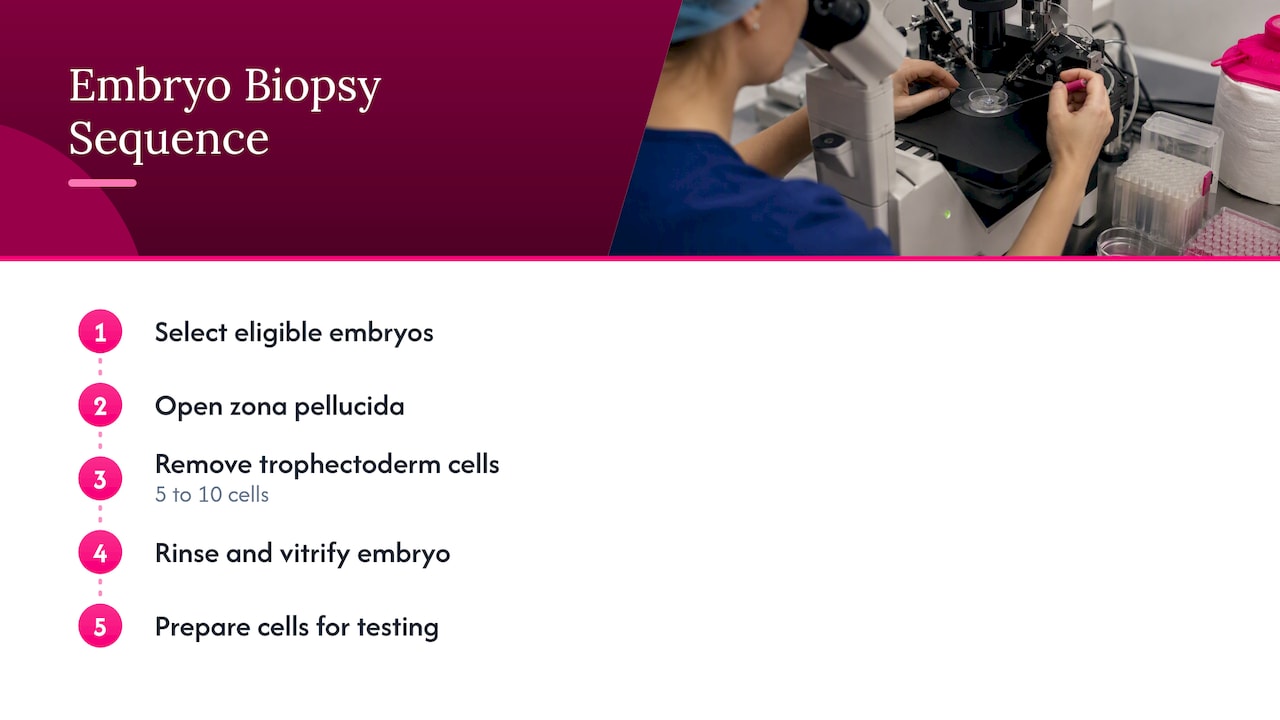
Step 1: Choosing which embryos to biopsy
The embryologist reviews each embryo’s development and appearance. Only embryos that have reached the right stage and quality are biopsied. Not every embryo from a cycle will qualify, and that is a normal part of the process.
Step 2: Opening the zona pellucida
Every embryo is surrounded by a protective shell called the zona pellucida. To reach the cells, the embryologist creates an opening in it, using a laser or a mechanical method. In the past, some labs used an acid solution for this step. The laser is the more common choice today. Research on the technique cautions that excess laser shots may induce a higher frequency of mosaicism, which is why careful, minimal laser use matters, as noted in a review of trophectoderm biopsy for PGT.
Step 3: Removing the cells
Using a very fine micropipette (a glass tube much thinner than a hair), the embryologist gently aspirates cells. At the blastocyst stage, 5 to 10 trophectoderm cells are removed. These cells belong to the outer layer that goes on to form the placenta, while the inner cell mass that becomes the baby is left alone. The ASRM committee opinion on PGT-A notes that trophectoderm biopsy is generally accepted to have less effect on embryo viability than cleavage-stage biopsy, because the removed cells do not have a fetal fate. You can read that in the ASRM 2024 committee opinion on PGT-A.
The UK regulator’s patient guidance adds a reassuring point: an embryo can still develop with fewer cells if the removal is done carefully, as stated on the HFEA page about PGT-A.
Step 4: Rinsing and freezing the embryo
The biopsied embryo is rinsed and cryopreserved (vitrified), as described by ESHRE. Freezing gives the genetics lab time to analyze the cells without rushing a transfer, and it allows your body to return to a natural state before an embryo is placed in the uterus.
Step 5: Preparing the cells for the genetics lab
The sampled cells are either fixed on a slide for a technique called FISH or, more commonly, collected in small reaction tubes for amplification-based testing. ESHRE reports that the tube method is the most widely applied. The DNA is copied (amplified) and then analyzed to produce a report for each embryo.
How long does an embryo biopsy take?
The hands-on part is short. A published review of trophectoderm biopsy states that the procedure should be performed within 3 minutes for each embryo. That time limit exists because embryos are kept outside their ideal culture conditions during the procedure, so labs work quickly and in a controlled environment.
The whole process from biopsy to results takes longer, because the cells must be shipped or transferred to a genetic testing lab and analyzed. Your clinic will give you its expected turnaround. Because the embryo is frozen while you wait, there is no pressure to complete a transfer within a fixed window.
What happens to the embryo and the cells afterward
The embryo stays frozen in storage until you and your care team choose it for a frozen embryo transfer. The cells go to the genetic analysis. When the report comes back, each embryo is typically classified in one of a few ways: no abnormality detected, abnormality detected, mosaic, or no result.
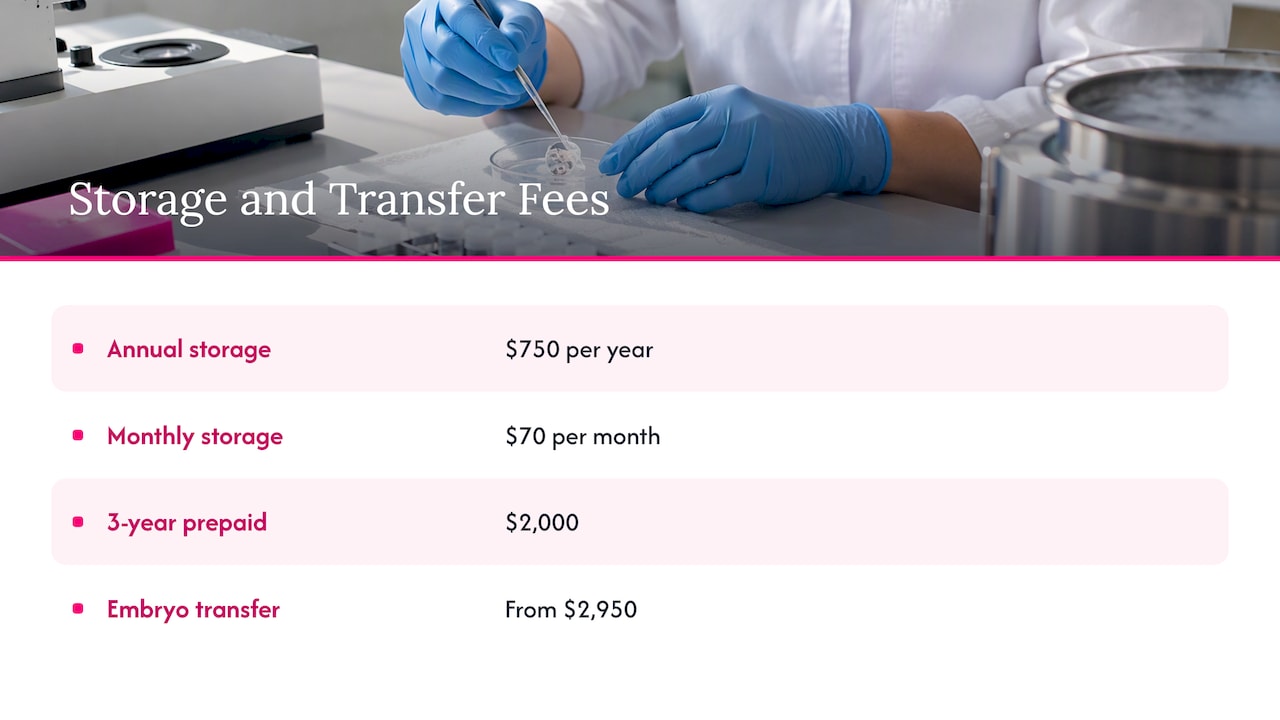
Storage is a practical consideration, since biopsied embryos wait in the freezer. At Life IVF Center, published storage fees are Annual Storage at $750 per year, Monthly Storage at $70 per month, or a 3 Year Prepaid option at $2,000. Embryo Transfer is listed from $2,950 on our treatment packages page.
Risks of embryo biopsy
Biopsy is an intervention on a living embryo, so it carries some risk, and a good clinic explains it before you consent.
- Damage to the embryo. The ASRM committee opinion says that while trophectoderm biopsy is considered safer than older cleavage-stage methods, risks remain, including potential damage to the embryo or blastocyst collapse. The review of trophectoderm biopsy adds that biopsy without assisted hatching may cause blastocyst collapse with a higher probability.
- Possible pregnancy-related associations. Some studies have suggested links between biopsy and a higher risk of hypertensive disorders of pregnancy. ASRM describes this evidence as limited and often confounded by patient factors.
- Long-term outcomes. A review of obstetric, neonatal and child health outcomes concludes that evidence on long-term consequences is inadequate, and that findings about biopsy are limited and may be confounded by other embryo manipulation or by patient characteristics. You can read the review of outcomes following embryo biopsy.
The practical takeaway is that technique and laboratory experience matter. Ask any clinic how many biopsies its embryologists perform, which method they use to open the zona, and whether the lab is on site. Keeping the lab in-house gives a clinic direct control over embryo handling from fertilization through freezing.
What biopsy results can and cannot tell you
PGT is informative, but it is not a perfect picture. The ESHRE paper and current clinical guidance point to several limits:
- Inconclusive or failed results. Sometimes the lab cannot produce a clear diagnosis. Rebiopsy can be considered in cases of failed, incomplete or inconclusive results, although the effect of a second biopsy on further embryo development is still under investigation.
- Mosaicism. A result can show a mix of normal and abnormal cells. Decisions about whether to transfer a mosaic embryo are individual and should be made with your physician and a genetic counselor.
- Sampling limits. The few cells removed may not perfectly represent the entire embryo.
- Scope. PGT does not identify all genetic risks. A normal result does not rule out every condition.
These limits are why we encourage patients to see PGT as one tool inside a broader plan, not a promise. Your age, ovarian reserve, medical history and the number of embryos available all affect whether biopsy is a good fit.
Our perspective
At Life IVF Center, we think about embryo biopsy as one decision inside a whole treatment plan, not as a routine add-on for every patient. Our Precision IVF® approach starts with your biology, history and goals, and only then asks whether genetic testing will give you information you can act on.
We see this most with patients who have a complex history: advanced maternal age, diminished ovarian reserve, endometriosis, immune disorders, a known genetic condition, or earlier cycles that did not go as hoped. For these patients, knowing more about each embryo can help shape which one to transfer first and how to plan next steps. Our in-house laboratory gives our team direct oversight of embryo culture, handling and freezing, which matters when the process involves a delicate procedure like biopsy.
We also believe you deserve a straight explanation of the limits. Results can be inconclusive, mosaic results require careful counseling, and no test guarantees a pregnancy. We welcome second-opinion cases, including patients who were told elsewhere that options were limited, and we publish many of our prices so that you can plan the financial side with confidence.
Questions to ask before you consent to a biopsy
Whether you are with us or comparing programs, these questions help you judge the quality of the process:
- At what stage do you biopsy, and why that stage for me?
- Is the embryology and freezing done in your own laboratory?
- What method do you use to open the zona pellucida?
- What happens if my results are inconclusive or mosaic? Is rebiopsy possible?
- Which type of PGT is right for my situation: PGT-A, PGT-M or PGT-SR?
- What are the costs for testing, freezing, storage and the transfer that follows?
A clear, unhurried answer to each is a good sign. If you are early in your planning, our pre-IVF testing price list shows the evaluations many patients complete first, such as Carrier Screening for Genetic Disease at $349 and Karyotype Chromosomal Analysis at $595.
Planning your cycle with Life IVF Center
If you are weighing PGT, the next step is a consultation where your history and test results are reviewed and a plan is built for you. We offer Precision IVF® at Life IVF Center, along with genetic testing, egg and embryo freezing, and donor and surrogacy pathways, all explained on our care overview. Our published packages include the Advanced IVF Package at $13,500, and you can see current details on the treatment packages page. Our team also supports patients with multi-language help, travel coordination and financial guidance.
If you want to compare freezing costs, Precision Embryo Freezing is listed at $8,950 on our freezing pricing page.
FAQ
Does an embryo biopsy hurt the embryo?
A biopsy is a real intervention, but it is designed to protect viability. At the blastocyst stage, the cells removed come from the layer that forms the placenta, not the part that becomes the baby, and professional guidance considers this less disruptive than cleavage-stage biopsy. Some risk of damage or blastocyst collapse remains, which is why experienced embryologists matter.
How long does an embryo biopsy take?
The procedure itself should take about 3 minutes per embryo, according to published technique reviews. The wait for genetic results is longer, and your clinic will tell you its typical turnaround. Embryos are frozen in the meantime.
Will I feel anything or need to rest afterward?
No. The biopsy is done on the embryo in the laboratory, not on your body, so there is no recovery from the biopsy itself. Your recovery needs relate to egg retrieval and, later, the embryo transfer, and your care team will give you instructions for each.
What if my biopsy results are inconclusive or mosaic?
An inconclusive result may lead to a rebiopsy, and a mosaic result may lead to a conversation about whether to transfer that embryo. Both are known limits of PGT. Your physician and a genetic counselor should walk you through your specific report.
Is embryo biopsy the same as an endometrial biopsy?
No. An embryo biopsy removes a few cells from an embryo in the lab for genetic testing. An endometrial biopsy samples tissue from the uterine lining, and it is a different procedure done for different reasons, such as an Endometrial Receptivity Assay.
Sources
- ESHRE PGT Consortium and SIG Embryology good practice recommendations for polar body and embryo biopsy
- Preimplantation genetic testing guidelines of the International Society of Reproductive and Developmental Medicine
- Preimplantation genetic testing: A narrative review
- Trophectoderm biopsy for preimplantation genetic testing review
- HFEA: Pre-implantation genetic testing for aneuploidy (PGT-A)
- Obstetric, neonatal, and child health outcomes following embryo biopsy
This article is for general educational purposes only and is not a substitute for personalized medical advice, diagnosis, or treatment. Fertility care is highly individual; please consult a qualified healthcare professional about your specific circumstances.
Ready to take the next step?
Life IVF Center specializes in individualized Precision IVF® care for complex cases—including diminished ovarian reserve, prior failed cycles, and advanced maternal age. Our in-house labs and dedicated physicians are ready to help.
Book a Free Consultation →