

A healthy embryo is defined by specific, measurable morphological features that predict its ability to implant and develop into a successful pregnancy. For couples and individuals going through IVF, understanding the signs of healthy embryo development is not abstract science. It is the foundation of every transfer decision your care team makes. Embryo quality assessment relies on cell number, symmetry, fragmentation levels, compaction timing, and blastocyst structure. Life IVF Center uses these clinical markers alongside advanced imaging technology to guide every step of the selection process.
1. Signs of a healthy embryo at Day 3
Day 3 is the first major checkpoint in embryo quality assessment. Embryologists assess embryo quality primarily by cell number, fragmentation, and symmetry at this stage to select the best embryos for IVF transfer.
A healthy Day 3 embryo shows three core features:
- Cell number: A healthy Day 3 embryo typically has 6–8 evenly sized cells. Fewer than 6 cells suggests slow development. More than 10 cells may indicate abnormal, accelerated division.
- Cell symmetry: Each cell, called a blastomere, should be roughly equal in size. Uneven blastomeres signal irregular division, which reduces implantation potential.
- Fragmentation: Fragmentation refers to small, non-cellular debris inside the embryo. Levels below 25% are acceptable. High fragmentation correlates with chromosomal abnormalities and lower live birth rates.
Cell number on Day 3 and degree of compaction on Day 4 are independent predictors of transferable blastocyst formation, surpassing fragmentation alone as predictors of success. That finding reframes how embryologists prioritize features during early assessment.
Embryos with well-distributed, mononucleated cells and minimal vacuoles tend to have higher live birth rates. Vacuoles are fluid-filled spaces inside cells that disrupt normal function when present in excess.
Pro Tip: Ask your embryologist for a written Day 3 morphology report. Knowing your embryo’s cell count and fragmentation percentage helps you understand the transfer decision and ask better questions before Day 5.
2. How morula compaction on Day 4 predicts viability
Day 4 is a milestone that many fertility patients never hear about, yet it carries strong predictive weight. Morula compaction is the process by which individual cells begin to merge into a tightly packed mass, losing their distinct borders. This happens between Day 4 and early Day 5.
Compaction matters because it signals that the embryo’s cells are communicating and adhering correctly. Without proper compaction, the embryo cannot form a blastocyst, which is the stage required for transfer or freezing.
The clinical distinction between full and partial compaction is significant:
- Full compaction: All cells are tightly merged with no visible cell borders. This is the strongest positive sign.
- Partial compaction: Some cells compact while others remain separate. Outcomes are intermediate.
- No compaction: Cells remain visibly distinct. The embryo is unlikely to progress to a viable blastocyst.
Top-quality embryos reaching full morula compaction on Day 4 show a live birth rate of 38.9% per transfer. That figure comes from a large cohort study of more than 3,100 blastocysts, making it one of the most statistically grounded benchmarks in current embryology.
Normal cleavage patterns and full morula compaction yield superior pregnancy and live birth outcomes compared to abnormal cleavage or partial compaction. Compaction status is now considered a critical selection criterion in protocols that integrate early cleavage timing, Day 3 morphology, and blastocyst quality.
3. What ICM and TE morphology reveal about embryo health
By Day 5, a healthy embryo becomes a blastocyst with two distinct structures. The inner cell mass (ICM) is the cluster of cells that will become the fetus. The trophectoderm (TE) is the outer layer that will form the placenta. Embryologists grade each structure separately on a scale from A (best) to C (poor).
ICM and TE grades independently predict chromosomal euploidy, with good grades correlating to a higher likelihood of chromosomally normal embryos. Euploidy means the embryo has the correct number of chromosomes, which is the single strongest predictor of successful implantation.
The practical implication is significant. Embryo morphology assessment correlates with euploidy rates, suggesting the potential to reduce reliance on invasive genetic testing by using ICM and TE grades. Preimplantation genetic testing for aneuploidy (PGT-A) is expensive and adds procedural risk. Morphology-based grading offers a non-invasive complement or, in some cases, an alternative.
| ICM Grade | TE Grade | Clinical Implication |
|---|---|---|
| A (tightly packed, many cells) | A (many cells, cohesive layer) | Highest euploidy likelihood |
| B (loosely grouped, several cells) | B (few cells, loose epithelium) | Moderate viability, acceptable for transfer |
| C (very few cells) | C (very few large cells) | Lower euploidy rate, transfer with caution |
Detailed separate evaluation of ICM and TE morphology is more informative than a combined score for predicting euploidy. Clinics that report a single blastocyst grade without separating ICM and TE are providing less actionable information than those that grade each structure independently.
Pro Tip: When reviewing your blastocyst report, look for two separate letter grades, one for ICM and one for TE. A report showing only one grade may be combining them, which gives you less information about your embryo’s true potential.
4. How AI and time-lapse imaging improve embryo assessment
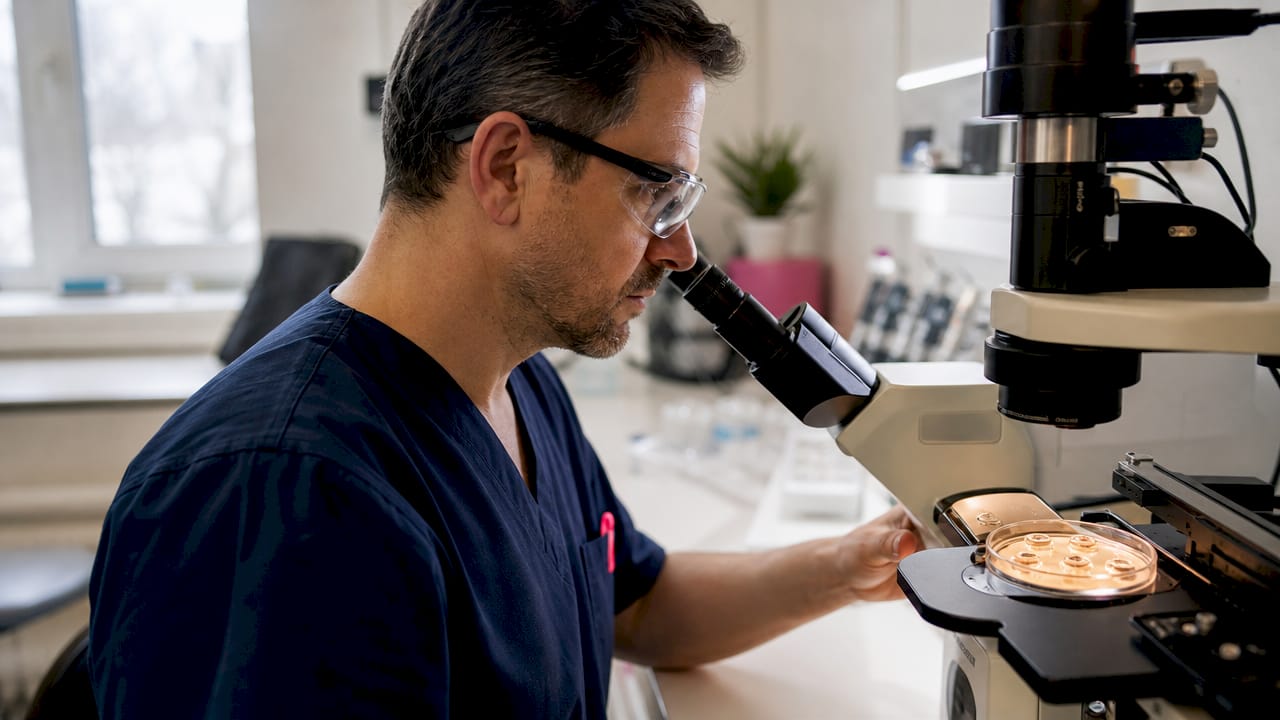
Traditional embryo grading depends on a single snapshot observation at fixed time points. That method introduces variability because different embryologists may score the same embryo differently. Time-lapse imaging addresses this by capturing continuous video of embryo development without disturbing the culture environment.
AI models using time-lapse embryonic imaging reduce variability in grading and improve prediction of implantation potential and euploidy status. The AI assessment accuracy in recent validation studies reached 72.5%, a meaningful improvement over purely subjective grading.
The benefits of AI-assisted assessment include:
- Consistency: AI applies the same criteria to every embryo, eliminating the inter-observer differences that affect manual grading.
- Continuous monitoring: Time-lapse systems record cell division timing, compaction onset, and blastulation without opening the incubator.
- Predictive depth: AI models detect subtle patterns in division timing that human observation misses, such as irregular cleavage intervals that predict chromosomal abnormality.
AI embryo assessment tools maintain predictive stability across datasets, enhancing clinical decision-making and optimizing resource allocation in IVF. External validation across multiple IVF clinics confirms that these tools perform consistently, not just in the labs where they were developed.
“Automation through AI reduces inter-observer variability in grading, enabling more consistent embryo viability assessment across clinics. This standardization is one of the most meaningful advances in IVF laboratory practice in the past decade.”
Life IVF Center integrates technology-driven IVF assessment into its Precision IVF® approach, combining morphological grading with AI-supported analysis to give each patient the most complete picture of their embryo’s potential. You can also learn more about the fertilization and culturing process to understand how embryos are monitored from the earliest stages.
Key Takeaways
Embryo selection accuracy depends on evaluating multiple independent markers across Days 3, 4, and 5, not on any single feature alone.
| Point | Details |
|---|---|
| Day 3 cell count matters | A healthy embryo has 6–8 evenly sized cells with fragmentation below 25%. |
| Morula compaction predicts live birth | Full compaction on Day 4 is linked to a 38.9% live birth rate per transfer. |
| Grade ICM and TE separately | Independent grading of each blastocyst structure predicts euploidy more accurately than a combined score. |
| AI reduces grading variability | Time-lapse AI tools reach 72.5% predictive accuracy and perform consistently across multiple clinics. |
| Multi-stage evaluation is best | Integrating Day 3 morphology, Day 4 compaction, and Day 5 blastocyst grades gives the strongest prediction of success. |
What I’ve learned about reading embryo quality reports
After years of observing how fertility patients process embryo quality information, one pattern stands out clearly. Patients who understand the difference between a Day 3 grade and a Day 5 blastocyst grade make better decisions and experience less anxiety during the wait between retrieval and transfer.
The most common mistake I see is treating a single morphology score as a verdict. A Day 3 embryo graded as “fair” can still develop into a high-quality blastocyst. Conversely, a “good” Day 3 embryo can arrest before Day 5. The full picture only emerges when you track the embryo across all three checkpoints: Day 3 morphology, Day 4 compaction, and Day 5 blastocyst structure.
The shift toward AI-assisted grading is real and meaningful, but it does not replace the embryologist. What AI does is remove the inconsistency that comes from human fatigue, lighting differences, and subjective interpretation. The embryologist still makes the final call, now with better data.
My practical advice: ask your clinic whether they use time-lapse imaging and whether ICM and TE are graded separately. If the answer to either question is no, ask why. The technology exists, the evidence supports it, and you deserve a complete assessment. Understanding your embryo transfer process in full is not just reassuring. It is the foundation of informed consent.
Personalized embryo assessment at Life IVF Center
Life IVF Center’s Precision IVF® approach builds embryo quality assessment into every stage of your treatment plan, from the first retrieval cycle through transfer selection.

The clinic’s Southern California laboratories use advanced morphological grading and AI-supported evaluation to assess each embryo across Day 3, Day 4, and Day 5 milestones. Every patient receives a personalized protocol based on their biological profile, not a standardized checklist. If you are ready to understand your embryo quality in full and work with a team that treats each case individually, review the fertility treatment packages at Life IVF Center to find the option that fits your clinical needs and budget. Consultations are available across multiple locations in Southern California.
FAQ
What does a healthy embryo look like on Day 3?
A healthy Day 3 embryo has 6–8 evenly sized cells and fragmentation below 25%. Cell symmetry and staged division are the primary quality indicators at this stage.
What is morula compaction and why does it matter?
Morula compaction is the process where embryo cells merge into a tight mass on Day 4. Full compaction is linked to a live birth rate of 38.9% per transfer, making it one of the strongest viability predictors in IVF.
What do ICM and TE grades mean on a blastocyst report?
ICM refers to the inner cell mass (future fetus) and TE refers to the trophectoderm (future placenta). Higher grades for each structure independently predict chromosomal normality and implantation success.
Can AI replace traditional embryo grading?
AI does not replace embryologists. It reduces grading variability and improves predictive accuracy to around 72.5%, giving clinicians better data to support transfer decisions.
How do I know if my embryo has a good chance of implanting?
The strongest indicators are full morula compaction on Day 4, a cell count of 6–8 on Day 3, and high ICM and TE grades on Day 5. Your embryologist should report all three checkpoints before a transfer decision is made.
Recommended
- Blog – Life IVF Center
- Understanding embryo transfer: The key step in IVF
- How to Support Your Partner Through IVF: A Complete Guide
- Egg retrieval tips: Expert guidance for IVF success
Ready to take the next step?
Life IVF Center specializes in individualized Precision IVF® care for complex cases—including diminished ovarian reserve, prior failed cycles, and advanced maternal age. Our in-house labs and dedicated physicians are ready to help.
Book a Free Consultation →