
Follicle monitoring, clinically known as folliculometry or a follicular study, is the medical process of using serial transvaginal ultrasounds to track ovarian follicle growth and confirm ovulation timing during a menstrual cycle. The role of follicle monitoring extends well beyond simple imaging. It reveals whether ovulation actually occurs, identifies disorders that hormone tests cannot detect, and gives fertility specialists the real-time data needed to time procedures like IUI and IVF with precision. For patients at Life IVF Center, this process forms a core part of individualized treatment planning, connecting biological events directly to clinical decisions.
How is follicle monitoring performed during the menstrual cycle?
Follicle monitoring follows a structured schedule tied to the menstrual cycle. Scans typically begin between days 8 and 11, then repeat every 1–3 days until the follicle reaches maturity. Most patients complete 3–6 ultrasound sessions per cycle.

Each session uses a transvaginal ultrasound probe to measure follicle diameter and assess the uterine lining. The sonographer records endometrial thickness and checks for a trilaminar (triple-line) pattern, which signals that the lining is receptive for implantation. Both measurements together give a fuller picture of cycle health than follicle size alone.
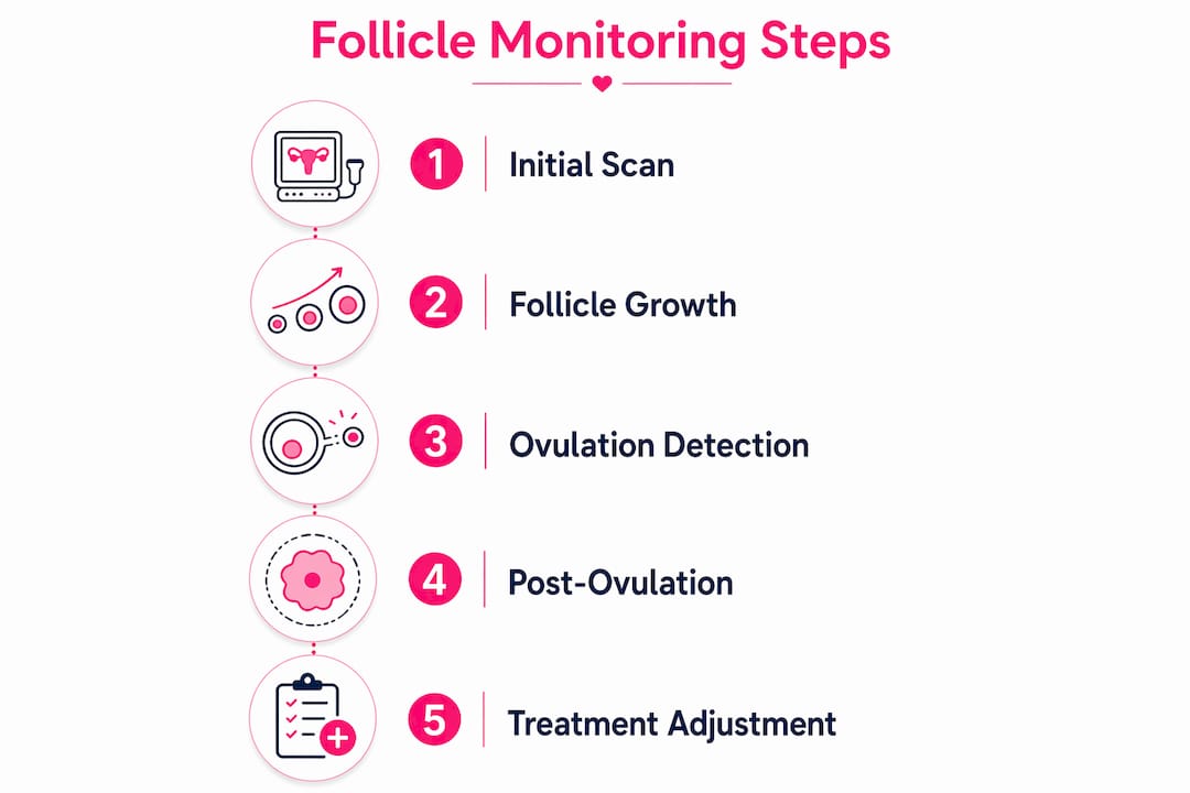
The sequence of a standard follicular study looks like this:
- Baseline scan (days 8–11): Identifies the dominant follicle and establishes the starting size.
- Intermediate scans (every 1–3 days): Tracks growth rate. A healthy dominant follicle grows approximately 1–1.5 mm per day toward a mature size of 18–24 mm.
- Pre-ovulation scan: Confirms the follicle has reached maturity and the endometrium shows adequate thickness.
- Post-ovulation scan: Verifies follicle collapse, free pelvic fluid, and corpus luteum formation at the rupture site, confirming that ovulation occurred.
The procedure itself is brief and well-tolerated by most patients. Discomfort is minimal, comparable to a standard pelvic exam. Costs vary by clinic and location, and patients should confirm coverage with their insurance provider before beginning.
Pro Tip: Patients with irregular cycles benefit most from anchoring scan timing to biological signs rather than calendar day counts. Combining follicle monitoring with a structured charting method like the Creighton Model reduces the risk of missing the fertile window due to cycle length variation.
What does follicle monitoring reveal about ovulation and fertility health?
Follicle monitoring provides a dynamic view of ovulation that no single hormone test can replicate. A blood draw captures one moment in time. Serial ultrasound captures the entire event, from follicle growth through rupture and corpus luteum formation.
The key diagnostic insights follicle monitoring delivers include:
- Ovulation confirmation: The three signs of confirmed ovulation are follicle collapse, the appearance of free pelvic fluid in the pelvis, and corpus luteum development at the rupture site.
- Mature follicle sizing: A follicle ready to ovulate measures approximately 18–24 mm. Follicles that fail to reach this size may indicate poor ovarian response.
- Luteinized Unruptured Follicle Syndrome (LUFS) detection: In LUFS, the follicle luteinizes and produces progesterone without actually releasing the egg. Hormone tests appear normal. Only serial ultrasound reveals that the follicle never collapsed.
- Endometrial assessment: Lining thickness below 7 mm or an absent trilaminar pattern suggests implantation may be compromised, even when ovulation is confirmed.
- PCOS and anovulation patterns: Patients with Polycystic Ovary Syndrome often show multiple small follicles that fail to develop a single dominant follicle. Monitoring tracks whether ovulation induction therapy produces a proper response.
A critical distinction separates follicle monitoring from an antral follicle count (AFC). AFC is performed on days 2–5 to estimate total ovarian reserve. Follicle monitoring is performed mid-cycle to track one follicle through its full growth and release. They answer different clinical questions and are not interchangeable.
Trained sonographers identify ovulation day with 84% accuracy using follicle collapse and corpus luteum criteria. That level of accuracy is clinically significant because it allows treatment timing to be anchored to a confirmed biological event rather than an estimate.
Pro Tip: A single mid-cycle hormone measurement can miss LUFS entirely. Serial scans over 3–6 sessions give a far more reliable picture of whether true ovulation is occurring.
| Observation | Clinical meaning |
|---|---|
| Follicle collapse + free fluid | Ovulation confirmed |
| Corpus luteum formation | Post-ovulatory phase confirmed |
| Follicle persists without collapse | Possible LUFS or anovulation |
| Endometrium below 7 mm | Implantation risk elevated |
| Multiple small follicles, no dominant | Consistent with PCOS pattern |
How does follicle monitoring support fertility treatment outcomes?
The benefits of monitoring follicles become most apparent when fertility treatment is involved. Precise timing is the single biggest variable in whether a cycle succeeds or fails, and follicle monitoring provides that precision directly.
The practical ways follicle tracking techniques improve treatment outcomes include:
- Timed intercourse: For patients attempting natural conception with timing difficulties, monitoring identifies the 24–48 hour window before ovulation when conception is most likely.
- IUI timing: Intrauterine insemination must be performed within a narrow window after the LH surge. Follicle monitoring confirms when the follicle is ready, reducing the risk of mistimed procedures.
- IVF stimulation monitoring: During IVF, patients take ovarian stimulation medications to grow multiple follicles. Serial scans track each follicle’s size and count, guiding the decision on when to trigger egg retrieval.
- Ovulation induction management: Medications like letrozole and clomiphene require monitoring to confirm they are producing an adequate response. Ultrasonographic follicular scoring can substitute for repeated estradiol blood draws in many patients, including those with PCOS.
- Ovarian hyperstimulation syndrome (OHSS) prevention: When scans show too many large follicles developing simultaneously, the physician can reduce medication dosage or delay the trigger shot to prevent OHSS, a potentially serious complication.
The financial case for monitoring is equally clear. Failed cycles carry both emotional and financial costs. Ultrasonographic follicular scoring reduces the burden of repeated fertility treatments by improving the accuracy of ovulation induction management. Fewer wasted cycles means lower cumulative costs and less emotional strain.
Follicle monitoring also integrates well with structured cycle charting. Combining monitoring with Creighton Model charting anchors scan timing to biological events rather than generic calendar days, which is especially valuable for patients with irregular cycles. You can review how follicle tracking fits into a broader fertility evaluation checklist to understand where it sits within a full diagnostic workup.
Pro Tip: No two cycles are identical. Follicle monitoring gives your care team the data to adjust medication doses, shift procedure timing, and respond to your specific cycle rather than a population average.
Who actually needs follicle monitoring, and what are its limits?
Follicle monitoring is not a universal requirement for every patient trying to conceive. Specialists recommend it primarily for patients with irregular cycles, ovulatory disorders, or those undergoing assisted reproductive technologies. Healthy patients with regular cycles and no known fertility issues typically do not need serial monitoring to conceive naturally.
The patients who benefit most from follicle development assessment include:
- Patients with irregular or unpredictable cycles who cannot reliably predict ovulation
- Patients with a diagnosis of PCOS, anovulation, or unexplained infertility
- Patients using ovulation induction medications like letrozole or clomiphene
- Patients preparing for IUI or IVF procedures
- Patients with a history of recurrent pregnancy loss where cycle quality is under investigation
Follicle monitoring does have real limitations. Results depend heavily on the timing of the first scan and the skill of the sonographer. A single scan without serial follow-up can be misleading. Monitoring also does not replace hormonal testing. Progesterone levels, LH measurements, and AMH values each provide information that ultrasound alone cannot supply.
Follicle monitoring is most powerful when it is one part of a coordinated fertility assessment, not a standalone test. Pairing it with hormonal data and clinical history gives the clearest picture of what is actually happening in a given cycle.
Many patients also assume that a normal-looking follicle guarantees ovulation. It does not. LUFS is the clearest example of why serial imaging matters. The follicle can appear healthy and reach mature size while still failing to release the egg. Understanding this distinction helps patients and clinicians avoid false reassurance. For a broader look at fertility misconceptions, the Life IVF Center resource on common fertility myths addresses several related misunderstandings that affect treatment decisions.
Key Takeaways
Follicle monitoring is the most direct method available for confirming ovulation, detecting ovulatory disorders, and timing fertility treatments with clinical accuracy.
| Point | Details |
|---|---|
| Monitoring confirms ovulation | Follicle collapse, free pelvic fluid, and corpus luteum formation together confirm ovulation occurred. |
| Serial scans outperform single tests | Repeated ultrasounds detect LUFS and anovulation that hormone tests alone cannot identify. |
| Best suited for specific patients | Monitoring is most valuable for irregular cycles, PCOS, ovulation induction, IUI, and IVF cases. |
| Supports treatment precision | Scan data guides medication dosing, procedure timing, and OHSS prevention in assisted reproduction. |
| Distinct from antral follicle count | AFC measures ovarian reserve early in the cycle; follicle monitoring tracks one follicle through ovulation. |
What I’ve learned from watching follicle monitoring change treatment decisions
After years of observing fertility care, the finding that still surprises patients most is how often a “normal” hormone panel coexists with a genuine ovulatory problem. LUFS is the clearest example. Progesterone rises, the patient believes ovulation occurred, and yet the egg was never released. Without serial ultrasound, that cycle would have been counted as a success and the underlying issue would have gone unaddressed for months.
The second pattern I see regularly is patients who arrive convinced they need monitoring for every cycle, regardless of their situation. Education matters here. A patient with a regular 28-day cycle, confirmed ovulation by temperature charting, and no history of fertility issues does not gain much from serial scans. Directing monitoring resources toward patients who genuinely need them produces better outcomes for everyone.
The most meaningful shift in recent years is the integration of follicle monitoring with individualized cycle data. When scan timing is anchored to a patient’s own biological markers rather than a generic day count, the results are more reliable and the treatment adjustments are more targeted. That kind of personalization is exactly what separates a well-designed fertility protocol from a one-size-fits-all approach. Patients who engage actively with their monitoring data, ask questions about what each scan shows, and work closely with their care team consistently report feeling more in control of a process that can otherwise feel opaque.
Follicle monitoring at Life IVF Center: personalized care from the first scan
Life IVF Center builds follicle monitoring into its fertility protocols as a standard component of individualized care, not an optional add-on. Whether you are preparing for IUI, moving through an IVF stimulation cycle, or investigating an ovulatory disorder, the clinical team uses serial scan data to adjust your protocol in real time.

The clinic’s Precision IVF® approach integrates follicle tracking with hormonal data and patient history to time every intervention to your specific cycle. Life IVF Center also offers fertility treatment packages designed to make monitoring-supported care accessible without unnecessary cost. If you are ready to understand what your cycle is actually doing, a consultation with the Life IVF Center team is the clearest next step.
FAQ
What is follicle monitoring in fertility treatment?
Follicle monitoring, also called folliculometry, uses serial transvaginal ultrasounds to track ovarian follicle growth and confirm ovulation during a menstrual cycle. It gives fertility specialists real-time data to time treatments like IUI and IVF accurately.
How many ultrasound scans does follicle monitoring require?
Most patients complete 3–6 scans per cycle, starting between days 8 and 11 and repeating every 1–3 days until ovulation is confirmed. The exact number depends on cycle length and how quickly the follicle matures.
Can follicle monitoring detect ovulation problems that blood tests miss?
Yes. Conditions like Luteinized Unruptured Follicle Syndrome produce normal hormone levels while the egg is never actually released. Serial ultrasound is the only reliable method to detect this type of ovulatory disorder.
Is follicle monitoring necessary for everyone trying to conceive?
Follicle monitoring is recommended primarily for patients with irregular cycles, ovulatory disorders, or those undergoing ovulation induction, IUI, or IVF. Patients with regular cycles and no known fertility issues typically do not require it for natural conception.
How does follicle monitoring differ from an antral follicle count?
An antral follicle count is a single scan performed on days 2–5 to estimate ovarian reserve. Follicle monitoring is a series of mid-cycle scans that track one dominant follicle through growth and release. They measure different aspects of reproductive health.
Recommended
- The Role of Stem Cells in Fertility: 2026 Guide
- Signs of Fertility Decline: What to Watch After 30
- Blog – Life IVF Center
- Your fertility evaluation checklist: Steps, tests, and benchmarks
Ready to take the next step?
Life IVF Center specializes in individualized Precision IVF® care for complex cases—including diminished ovarian reserve, prior failed cycles, and advanced maternal age. Our in-house labs and dedicated physicians are ready to help.
Book a Free Consultation →